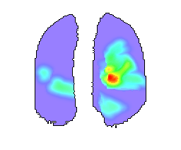
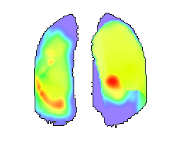
Abstract: Here we present the first version of a statistical atlas of lung lesions. The lesions considered in this study are of different nature and different biological substrates. Despite these lesion types are very common, they may be partly associated with lung tuberculosis because they were detected, isolated, and segmented on lung CT images of tuberculosis (TB) patients. The ultimate goal of this study and related free electronic resources is to provide the relevant part of the society for 3D statistical maps of the frequency of lung lesions of different kinds.


Reference: Kovalev V., Liauchuk V., Gabrielian A., Rosenthal A. Towards Statistical Atlas of Lung Lesions. International Journal of Computer Assisted Radiology and Surgery, vol. 15, Suppl. 1, 2020 [in press].
Resources
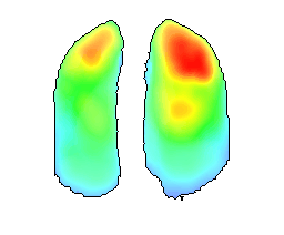
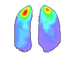
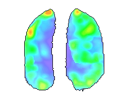
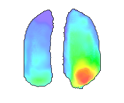
Previews of 3D atlases for six different types of lesions are shown below.
Click Download All to download all the resources at once.
The resources include 3D statistical data in Nifti file format along with some animated GIFs. Non-negative voxel values in Nifti files correspond to averaged frequency of occurrence of the corresponding type of lesion in every given anatomical location. Negative voxel values correspond to non-lungs region. Voxel size corresponds to 2.7 mm in all dimensions.
A freely-available tool “VV” can be used for viewing the Nifti image files.